Wednesday, April 15, 2015

Old School DNA Sequencing: Sanger Method


Before modern technology was available, DNA sequencing was much more tedious and difficult. In the 1950's Dr. Frederick Sanger determined the sequence of amino acids in protein.  This led to the understanding that DNA sequencing was collinear to the sequencing of amino acids. In the 1970's, Dr. Sanger developed a method to determine the exact sequence of nucleotides in a given gene. This method involved placing the DNA sample in  gel that was charred both negatively and positively. The longer strands of DNA would be pulled through the gel slower than the shorter strands, and the DNA separated into bands. The band positions were then "read" in order to determine DNA sequence. To stimulate this, we were given a lab packet in which DNA from four different subjects were tested. We were asked to read each sequence, compare them, and finally determine which subjects carried a disease. Pictures from the lab are posted below. 


In the step above, we determined the sequencing of each subject's DNA by evaluating the bands. This data was then placed in the chart pictured in the last image. 


After comparing the data, it became clear that "Norm" was the only healthy patient. All other subjects showed some disturbance in their DNA that caused them to be effected by the disease. Carol experienced a front shift mutation, Bob experienced a truncation mutation shift, and Abby experienced point mutation. This disease would be unrecognizable were it not for the Sanger Method.

Wednesday, April 8, 2015

DNA Extraction Lab

As we learn more about DNA, it is great to apply the concepts we learn so that we can gain an even deeper understanding. In this post, I am going to describe a lab we did in class in which we extracted DNA from wheat germ. In order to do this, our instructor had us work in groups so that we could collaborate our knowledge.

To begin the lab, we measured out 1 gram of raw wheat germ. This germ was then placed in a 50 ml test tube where it was mixed with 20 ml of hot tap water for 3 minutes. After being stirred, 1/4 teaspoon of detergent was added to the mixture. This was again stirred. 5 minutes after adding the detergent, we added 14 ml of alcohol. This was done very carefully so that the alcohol would not mix with the germ mixture. This was necessary in the extraction process, as DNA precipitates at the water-alcohol interface. The image below shows this step being done.


After doing this, the test tube had to sit for a few minutes so that the DNA could precipitate. After a short wait, we collected the DNA by using a wooden stick.


Overall, this was a really fun lab that expanded my knowledge of DNA.

Monday, April 6, 2015

From DNA to Protein: The Central Dogma

As we are aware, DNA is found in protein, but how does it get there? After all, DNA is located in the nucleus of all cells, and ribosome build protein. In order to cross the nucleus membrane in order for protein to be made, there has to be a step in-between that will transfer the DNA code to the ribosome, but what is that step? The answer lies in the process of the central dogma, and the material RNA. RNA, as you can predict, is very similar to DNA. Instead of having dioxyribose material, like in DNA, RNA contains ribose. As we will learn, RNA will be the key component to the central dogma, allowing protein to be successfully made. For now, we are going to define a few terms related to this process. To view the vocabulary, please click here.

Wednesday, April 1, 2015

DNAi Timeline

Like most things, DNA would not be as well understood as it is today without the contributions from multiple researchers. This post is going to detail the work from some of these contributing scientists that helped develop the current understanding of DNA. A great resource for information on other scientists that contributed work to the evolution of the idea of DNA can be found at this address. This is also the resource that was used for this post.

Martha Chase, Alfred Hershey, and the "Blender Experiment"
In the early 50's, a group of researchers at the Cold Spring Harbor Laboratory were researching bacteriophage genetics (also referred to as phage genetics). Phage are viruses that target, attack, and infect bacteria. At this time, it was already known that phage was constructed with an outer casing of protein that surrounded in inner core of DNA. It was also known that phage relied on bacteria to reproduce, and that phage attached themselves to bacteria by their tails. It was hypothesized that after attaching, the phage pumped genetic material into the bacterial host, causing the bacterial enzymes to be replaced by new phage particles. In 1952, Martha Chase and Alfred Hershey dedicated their work to discovering why the bacteria transformed into a phage producing organism, proving whether or not DNA was a transforming principle. Through chemical analysis, it was discovered that DNA contained high amounts of phosphorus and zero amounts of sulfur. This information was in contrast to the known information about protein, which contains sulfur but no phosphorus. This information was used in an experiment to test which component entered the bacteria for infection. This was designed in a way that radioactive phosphorus (32P) and radioactive sulfur (35P) to selectively label the phage DNA and protein. The radio labeled phage was then combined with unlabeled bacteria so that the phage could attach. The attachment was then disrupted as the culture was mixed in a Waring blender. The samples were then spun in a centrifuge in an attempt to separate the phage from the bacteria. This attempt was successful, and due to the fact that the phage is lighter than the bacteria, it remained suspended in the test tube while the bacteria collected at the bottom in the form of a pellet. After evaluating the sample 35P, it was discovered that the newly produced phage from the bacteria did not contain any radioactive sulfur, unlike the protein coat of the parent phage.  In contrast, the sample 32P contained newly produced phage from the bacteria that was contaminated with radioactive 32P. From this, it was determined that phage DNA was used inside the bacteria to produce new phage, confirming that DNA is the genetic material.

Monday, March 30, 2015

DNA Sketch Model

As we work further into the human body and system, we must work to understand one of the most key components to human function: DNA structure and function. To do this, we were given instruction about the basic components to DNA and asked to fit them together in a sketch that would demonstrate how it fit together to form a strand of DNA. The pictures included below are the "finished product" of this activity.

As you can see in the last picture, a nucleotide is shown. A nucleotide is the basic component of DNA, composed of dioxyribose, phosphate, and one Guanine, Cytosine, Adenine, or Thymine. Notice the "or" in that list. A nucleotide only contains one of the four listed, and can be found int the acronym GCAT. These then form a hydrogen bond with the opposite strand; the only possible combinations of pairings are Guanine with Cytosine, and Adenine with Thymine. Once the strands are bonded, they align themselves in a double helix shape, forming the common DNA perception. There you have it: the basics of DNA!


Thursday, March 12, 2015

Pedigrees and Genetic Counseling: In Sickness and Health

For this post, we are going to create a family pedigree for a couple who wishes to have children. We will evaluate the pedigree to predict the risk of their offspring having Hemophilia or Myotonic Dystrophy. We are also going to evaluate this situation to further understand the reasoning behind the risk prediction.

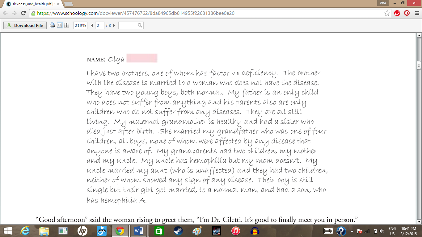
Shown below are the letters that Greg and Olga sent in regarding their families.


To set a basic understanding for this pedigree, I am going to define a few symbols shown. Each circle on the pedigree represents a female family member, while each square represents a male family member. Shapes completely filled in with purple represents family members affected by Hemophilia, while shapes filled in completely with blue represent family members affected by Myotonic Dystrophy. Shapes that are not colored in represent family members who are unaffected by either disorder.  Shapes that have a large X threw them represent deceased family members. The pedigree outlined in red represents Greg's side of the family while the pedigree outlined in green represents Olga's family.


Myotonic Dystrophy is classified as an"autosomal dominant disorder", meaning that a disease can be inherited through only one parent, if the gene is dominant. This means that the disorder is passed down directly, without skipping generations. It also means that if the disorder is not inherited by a child, it can no longer be passed down through that child. Because  Myotonic Dystrophy is autosomal dominant disorder, and the gene is not present in either Greg or Olga, they are not carriers of the gene and cannot pass the disorder down to their offspring.

When viewing the pedigree, on might wonder about the possibility of Greg's aunt or uncle being homogeneous for the Myotonic Dystrophy gene. Being that only one parent carried the gene, only one abnormal gene was inherited, causing the aunt and uncle to be heterogeneous for the Myotonic Dystrophy gene. You might also question how it can be determined that Greg's cousin does not have Myotonic Dystrophy, being that symptoms sometimes don't appear until after the age of fifty. Being that neither of the cousin's parents were affected by this gene, it can be determined that it would be impossible for the cousin to have Myotonic Dystrophy.

Now, let's discuss the matter of Hemophilia inheritance. As discussed, Myotonic Dystrophy is classified as an "autosomal dominant disorder", but what about Hemophilia?

Another type of classification of gene inheritance is that of an "autosomal recessive disorder", meaning that two abnormal genes are required in order to be affected by the disorder. If a person only inherits one abnormal gene, they become carriers of the disorder, however, they are not affected. This is the type of disorder that has the ability to skip generations. Is Hemophilia an autosomal recessive disorder?

It is important that we discuss another concept, that of consanguinity. Consanguinity is simply where two individuals from the same biological background mate. Consanguinity increases the probability of an individual involved to become a carrier of an autosomal recessive disorder. Again, this recessive trait can skip generations until it has the opportunity to create offspring that is homogeneous with the autosomal recessive disorder gene.

 When tracing the pattern of inheritance of the Hemophilia disorder, we begin to see a pattern. It becomes clear that only male individuals within the family inherited this disorder.  Hemophilia is a sex-linked disorder, indicating that Hemophilia is not an autosomal recessive disorder.

Sex-linked disorders are often classified in X-linked recessive inheritance. This means that specific disorders, such as Hemophilia, are attached to the X chromosome. Due to the fact that males do not pass down the X chromosome directly to their male offspring, the X-linked recessive inheritance is passed down through the female offspring, then on to the male grandchildren. Males do not receive the X chromosome from their father because the father passes down the Y chromosome down to the son while the mother passes down the X chromosome. The fathers can, however, pass down their X chromosome to their female offspring. This causes men to more likely be affected by diseases passed down through X-linked recessive inheritance. In order for females to display a sex-linked trait such as Hemophilia, she must inherit a defective X chromosome from both parents.

Why don't we apply the concept of carrying an  X-linked recessive gene to the pedigree of Greg and Olga? If we evaluate the family's history, we can see that Olga's mother, grandmother, and cousin were most definitely carriers of Hemophilia, while there is only a possibility of Olga being a carrier. If we evaluate Greg's family, we can see that Greg's mother, grandmother, and great-grandmother were definitely carriers while there is only the possibility of Greg's aunt and cousin being carriers. Greg is unaffected. Being that Olga has the possibility of being a carrier, there is a 50% chance that this trait would be passed down to her offspring. If the trait was passed down to a son, the son would be affected by the disorder. If the trait was passed down to a daughter, the daughter would only be a carrier. Again, there is only a 50% chance of Olga being a carrier, which means that there is the possibility that the trait will not be passed down to the offspring under any circumstance.

Human Chromosomes and Genetic Diseases

In this post, I am going to discuss how genetic diseases are inherited through human chromosomes while investigating some specific diseases. Before we begin, it is important to understand the basics of chromosomes. We have already discussed chromosomes frequently in previous posts, however, we have not yet labeled the specific components as of yet, which is very helpful in understanding genetics.
So how are genetic diseases actually linked to chromosomes? Do better understand this, we are going to look at a specific chromosome (such as the X chromosome shown above) and investigate some diseases attached. 

To begin with, we are going to research Rett Syndrome. This disease is attached to the X chromosome through the mutated gene MeCP2.  It is most common for this mutation to occur on X chromosomes transferred by sperm cells. Rett syndrome is a progressive neurodevelopmental disorder that affect females and very rarely affect males. A possible explanation as to why this disease is rare in males is that males do not inherit the X chromosome from their father, as girls do. As previously stated, most mutated MeCP2 genes are attached to the X chromosome inherited from the father. Rett syndrome occurs in about 1 of 10,000 births and is a common cause of mental impairment. This disease causes multiple symptoms in addition to mental impairment, such as the loss of hand usage ability, reduced muscle tone, seizures, intense eye gaze, and an "autistic-like" phase.    

Another disease we are going to look at is Alport Syndrome, which is also attached to the X chromosome. As MeCP2 was effected in Rett Syndrome, COL4A5 is mutated in order to develop Alport Syndrome. Alport Syndrome is a collagen-related disease affecting the kidneys, ears, and eyes.  Due the fact that the mutated gene associated with Alport Syndrome is passed along with the X chromosome, and the fact that women have two X chromosomes, affected women usually have one normal X chromosome, causing the effects to be much less drastic. This also means that since males only have on X chromosome, affected males will have more severe effects. Symptoms of Alport Syndrome include the breakdown of filters associated with glomerular basement membranes, causing the loss of proteins and red blood cells into the urine. Alport Syndrome also causes progressive deafness and issues regarding the lens of the eyes. 

Resources used for this post are listed below: